
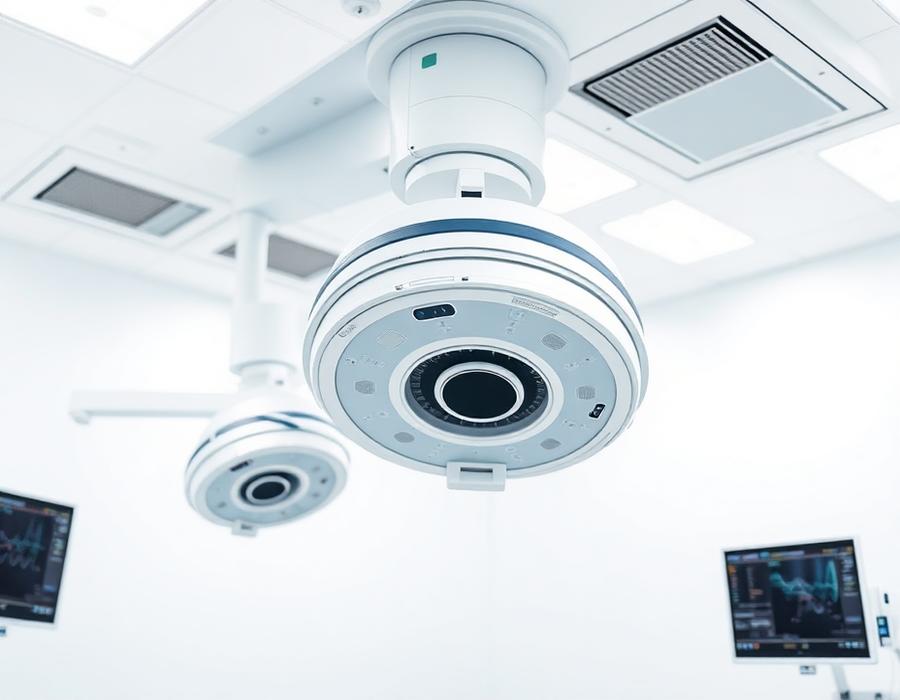
What a C-arm does
A C-arm provides real-time fluoroscopic imaging during a procedure. The C-shaped arm carries an X-ray source at one end and an image receptor at the other, allowing the surgeon to view anatomy and instrument position without moving the patient.
Mobile versus fixed
Mobile C-arms are wheeled between theatres and suit orthopaedics, trauma, general surgery, pain management and endourology. They are the pragmatic choice for facilities with variable theatre scheduling.
Fixed C-arms are installed in a dedicated room with higher output, better heat management and larger detectors. They are appropriate for high-volume interventional programmes running long procedures.
Image intensifier or flat panel
Image intensifier systems remain cost-effective and are widely supported. Flat panel detectors give distortion-free images across the whole field, better low-contrast visibility and a more compact profile, at a higher price point.
Specification points that matter
Generator output and whether the unit is pulsed; heat unit capacity for long procedures; field-of-view sizes; free space and orbital and angular movement ranges; last-image-hold, road-mapping and DSA if vascular work is planned; and DICOM connectivity to your archive.
Match the system to theatre volume
As a rule of thumb, occasional orthopaedic use is well served by a compact mobile unit; a busy multi-theatre orthopaedic and endourology programme benefits from a higher-output mobile system with a flat panel; and a dedicated cardiac or vascular interventional list needs a fixed installation.
This guide is educational only. Specifications, availability and regulatory status are confirmed per quotation and per unit.